SDAP Home Page
SDAP Overview
SDAP All
SDAP Food
Use SDAP All
Search SDAP All
List SDAP All
SDAP Food
SDAP Tools
FAO/WHO Allergenicity Test
FASTA Search in SDAP
Peptide Match
Peptide Similarity
Peptide-Protein PD Index
Aller_ML, Allergen Markup Language
List SDAP
About SDAP
General Information
Manual
FAQ
Publications
Who Are We
Advisory Board
Allergy Links
Our Software Tools
MPACK
FANTOM
GETAREA
NOAH/DIAMOD
MASIA
PCPMer
InterProSurf
EpiSearch
Protein Databases
PDB
MMDB - Entrez
SWISS-PROT
NCBI - Entrez
PIR
Protein Classification
CATH
CE
FSSP
iProClass
ProtoMap
SCOP
TOPS
VAST
Bioinformatics Servers
@TOME
BLAST @ NCBI
BLAST @ PIR
FASTA @ PIR
Peptide Match @ PIR
ClustalW @ BCM
ClustalW @ EMBL - EBI
ClustalW @ PIR
Bioinformatics Tools
Cn3D
MolMol
Bioinformatics Links
Bioinformatics Links Directory
|
SDAP - All Allergens
| Allergen Ara h 1Translate to AllerML | | | Allergen | Ara h 1 | Type | foods | Species - Systematic Name | Arachis hypogaea | Species - Common Name | peanut | Keywords | Vicilin; Clone P17
| Class | IUIS |
| |
Ara h 1 - PubMed | Reference | Reference 1 | Burks AW, Cockrell G, Stanley JS, Helm RM, Bannon GA. Recombinant peanut allergen Ara h I expression and IgE binding in patients with peanut hypersensitivity.J Clin Invest. 1995 Oct;96(4):1715-21. | Reference 2 | Kolarich D, Altmann F. N-Glycan analysis by matrix-assisted laser desorption/ionization mass spectrometry of electrophoretically separated nonmammalian proteins: application to peanut allergen Ara h 1 and olive pollen allergen Ole e 1. Anal Biochem. 2000 Oct 1;285(1):64-75. |
Ara h 1 - Protein Sequences FASTA@SDAP: FASTA search against all SDAP allergen sequences performed at SDAP BLAST@ExPASy: BLAST search performed at the Expert Protein Analysis System (ExPASy) proteomics server of the Swiss Institute of Bioinformatics (SIB) PROSITE@PIR: PROSITE search performed at PIR - Protein Information Resources
Ara h 1 - Protein Sequence Properties PeptideCutter@ExPASy: Protease cleavage sites predicted with PeptideCutter from the Expert Protein Analysis System (ExPASy) proteomics server of the Swiss Institute of Bioinformatics (SIB)
Ara h 1 - Protein Similarity, Classification and Clustering
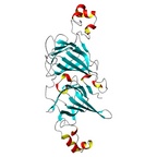
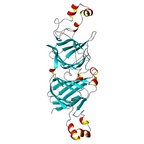
Ara h 1 - Protein Models These 3D allergen models are superseded by newer PDB structures.
Ara h 1 - Pfam domains Pfam is a database of multiple sequence alignments and hidden Markov models covering protein domains and families
Inspect MotifMate results for Ara h 1. AutoMotif contains a database of PCPMer motifs for allergens. MotifMate home page MotifMate summary Browse MotifMate proteins Browse MotifMate families
Ara h 1 - Sequences from the Entrez Nucleotides database
Link to GenBank | View Sequence | 602435 | Go! |
Ara h 1 - PDB Structures
PDB Accession | Jmol | Deposition date | Experimental | Observation | 3S7E | View! | 2011-05-26 | X-ray diffraction resolution 2.71 A | | 3S7I | View! | 2011-05-26 | X-ray diffraction resolution 2.35 A | | 3SMH | View! | 2011-06-28 | X-ray diffraction resolution 2.43 A | crystal structure of the major peanut allergen Ara h 1 |
Ara h 1 - EpitopesNo | Epitope | Exact Match @SDAP | Peptide Similarity @SDAP | Exact Match @PIR | Position | Type | Description | 1 | AKSSPYQKKT | Go! | Go! | Go! | 25-34 | IgE | When altered, amino acids shown in red lead to loss of IgE binding:
AKSSPYQKKT
| 2 | QEPDDLKQKA | Go! | Go! | Go! | 48-57 | IgE | When altered, amino acids shown in red lead to loss of IgE binding:
QEPDDLKQKA
| 3 | LEYDPRLVYD | Go! | Go! | Go! | 65-74 | IgE | When altered, amino acids shown in red lead to loss of IgE binding:
LEYDPRLVYD
Mutation: C71L
| 4 | GERTRGRQPG | Go! | Go! | Go! | 89-98 | IgE | When altered, amino acids shown in red lead to loss of IgE binding:
GERTRGRQPG
| 5 | PGDYDDDRRQ | Go! | Go! | Go! | 97-106 | IgE | When altered, amino acids shown in red lead to loss of IgE binding:
PGDYDDDRRQ
| 6 | PRREEGGRWG | Go! | Go! | Go! | 107-116 | IgE | When altered, amino acids shown in red lead to loss of IgE binding:
PRREEGGRWG
| 7 | REREEDWRQP | Go! | Go! | Go! | 123-132 | IgE | When altered, amino acids shown in red lead to loss of IgE binding:
REREEDWRQP
| 8 | EDWRRPSHQQ | Go! | Go! | Go! | 134-143 | IgE | When altered, amino acids shown in red lead to loss of IgE binding:
EDWRRPSHQQ
| 9 | QPRKIRPEGR | Go! | Go! | Go! | 143-152 | IgE | When altered, amino acids shown in red lead to loss of IgE binding:
QPRKIRPEGR
| 10 | TPGQFEDFFP | Go! | Go! | Go! | 294-303 | IgE | When altered, amino acids shown in red lead to loss of IgE binding:
TPGQFEDFFP
| 11 | SYLQEFSRNT | Go! | Go! | Go! | 311-320 | IgE | When altered, amino acids shown in red lead to loss of IgE binding:
SYLQEFSRNT
Mutation: G315E
| 12 | FNAEFNEIRR | Go! | Go! | Go! | 325-334 | IgE | When altered, amino acids shown in red lead to loss of IgE binding:
FNAEFNEIRR
| 13 | EQEERGQRRW | Go! | Go! | Go! | 344-353 | IgE | When altered, amino acids shown in red lead to loss of IgE binding:
EQEERGQRRW
| 14 | DITNPINLRE | Go! | Go! | Go! | 393-402 | IgE | When altered, amino acids shown in red lead to loss of IgE binding:
DITNPINLRE
| 15 | NNFGKLFEVK | Go! | Go! | Go! | 409-418 | IgE | When altered, amino acids shown in red lead to loss of IgE binding:
NNFGKLFEVK
| 16 | RRYTARLKEG | Go! | Go! | Go! | 498-507 | IgE | When altered, amino acids shown in red lead to loss of IgE binding:
RRYTARLKEG
| 17 | ELHLLGFGIN | Go! | Go! | Go! | 525-534 | IgE | When altered, amino acids shown in red lead to loss of IgE binding:
ELHLLGFGIN
| 18 | HRIFLAGDKD | Go! | Go! | Go! | 539-548 | IgE | When altered, amino acids shown in red lead to loss of IgE binding:
HRIFLAGDKD
| 19 | IDQIEKQAKD | Go! | Go! | Go! | 551-560 | IgE | When altered, amino acids shown in red lead to loss of IgE binding:
IDQIEKQAKD
| 20 | KDLAFPGSGE | Go! | Go! | Go! | 559-568 | IgE | When altered, amino acids shown in red lead to loss of IgE binding:
KDLAFPGSGE
| 21 | KESHFVSARP | Go! | Go! | Go! | 578-587 | IgE | When altered, amino acids shown in red lead to loss of IgE binding:
KESHFVSARP
| 22 | QNLRVAKISMPVNTPGQF | Go! | Go! | Go! | 281-298 | mAb-4A8 | The 4A8 MAb bound linear peptides 18, 29, 33 and 34:
| 23 | PHFNSKAMVIVVVNKGTG | Go! | Go! | Go! | 446-463 | mAb-4A8 | The 4A8 MAb bound linear peptides 18, 29, 33 and 34:
| 24 | EGDVFIMPAAHPVAINAS | Go! | Go! | Go! | 506-523 | mAb-4A8 | The 4A8 MAb bound linear peptides 18, 29, 33 and 34:
| 25 | ASSELHLLGFGINAENN | Go! | Go! | Go! | 521-538 | mAb-4A8 | The 4A8 MAb bound linear peptides 18, 29, 33 and 34:
| 26 | LVIQQGQATVTVANGNNR | Go! | Go! | Go! | 236-253 | mAb-13A4 | The 13A4 MAb bound linear peptides 15, 28 and 29
| 27 | MMLTCVEIKEGALMLPHF | Go! | Go! | Go! | 431-448 | mAb-13A4 | The 13A4 MAb bound linear peptides 15, 28 and 29
| 28 | PHFNSKAMVIVVVNKGTG | Go! | Go! | Go! | 446-463 | mAb-13A4 | The 13A4 MAb bound linear peptides 15, 28 and 29
|
1. D. S. Shin, C. M. Compadre, S. J. Maleki, R. A. Kopper, H. Sampson, S. K. Huang,
A. W. Burks, and G. A. Bannon,
Biochemical and structural analysis of the IgE binding sites on Ara h 1,
an abundant and highly allergenic peanut protein,
J. Biol. Chem. 1998, 273, 13753-13759.
2. Hnasko RM, Lin AV, McGarvey JA, Mattison CP.
Sensitive and selective detection of peanut allergen Ara h 1 by ELISA and lateral flow immunoassay.
Food Chem. 2022 Dec 1;396:133657. doi: 10.1016/j.foodchem.2022.133657. Epub 2022 Jul 8. PMID: 35843000.
3. Burks AW, Shin D, Cockrell G, Stanley JS, Helm RM, Bannon GA. Mapping and mutational analysis of the IgE-binding epitopes on Ara h 1, a legume vicilin protein and a major allergen in peanut hypersensitivity. Eur J Biochem. 1997 Apr 15;245(2):334-9. doi: 10.1111/j.1432-1033.1997.t01-1-00334.x. PMID: 9151961.
|